MED-TECH | Scientists resort to machine learning’s seeing powers on cell structures
Scientists at the Allen Institute used machine learning to train computers to see more clear parts of the cell, without the costly fluorescent labels.
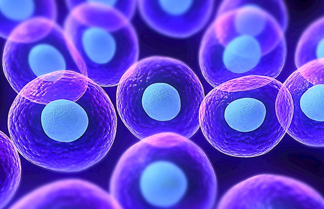
Conceptual cells
WASHINGTON — Scientists at the Allen Institute used machine learning to train computers to see more clear parts of the cell, without the costly fluorescent labels.
The study published on Monday in the journal Nature Methods described the AI technique that used only black and white images generated by inexpensive technique known as brightfield microscope. It allowed scientists to see larger picture of human cells that have more than 20,000 proteins.
“This technology lets us view a larger set of those structures than was possible before,” said Greg Johnson, scientist at the Allen Institute for Cell Science and the senior author on the study.
“This means that we can explore the organization of the cell in ways that nobody has been able to do, especially in live cells,” said Johnson.
Fluorescence microscopy, which uses glowing molecular labels to pinpoint specific parts of cells, is very precise but only allows scientists to see a few structures in the cell at a time.
The prediction tool could also help scientists understand what goes wrong in cells during disease, according to Rick Horwitz, Executive Director of the Seattle-based Allen Institute for Cell Science.
Cancer researchers could potentially apply the technique to archived tumor biopsy samples to better understand how cellular structures change as cancers progress or respond to treatment.
The algorithm could also aid regeneration medicine by uncovering how cells change in real time as scientists attempt to grow organs or other new body structures in the lab.
The researchers used an existing machine learning technique, known as a convolutional neural network, to train computers to recognize finer details in the black and white images, such as the mitochondria, cells’ powerhouses.
They tested 12 different cellular structures and the model generated predicted images that matched the fluorescently labeled images for most of those structures, according to the researchers.